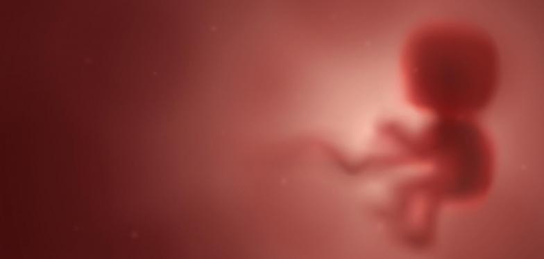
Angiography used to facilitate post-mortem examinations of foetuses
UCLouvain's Saint-Luc University Hospital has a Belgian first to its name. Injecting a specific, fat-absorbing contrast medium allows doctors to obtain extremely detailed and true-to-life imaging of the vascular system of a foetus that has died in utero, after a miscarriage or as a result of an abortion, thereby sparing parents the pain of a conventional autopsy.
Medical imaging by angiography, that is the mapping of blood vessels using a contrast medium, has been used for years in adults to detect abnormalities or lesions. But once death has set in, a liquid that mixes with oil is needed so that the results can still be seen. Saint-Luc has now succeeded in doing this in foetuses, which represents a remarkable advance. That is because foetuses are very small and because accurate dissection is sometimes impossible. In utero maceration and certain changes after death also make tissue analyses somewhat more difficult. Thanks to this specific form of angiography, many questions regarding the cause and circumstances of death can be clarified without the need to dissect the foetus.
This technique offers a whole variety of benefits. The injection is carried out via the umbilical cord and is therefore less invasive than injection via the thigh. The parents can be provided with valuable information about the cause of death of the foetus, they are also more likely to consent to an examination of this type as opposed to a conventional autopsy and all radiological data are stored.